Medical News Blog Information

Transfusion Medicine in Africa: Johannesburg, South Africa

James Kelley, PhD, MD
Department of Pathology
Brigham and Women�s Hospital

Like most visitors to southern Africa, my first stop goes through Johannesburg � the �city of gold�. This megalith of 7 million people conjures images of apartheid and armed home invasions; however, the people are as friendly and warm as its summer weather. After all, this is the home of Nelson Mandela and the World Cup. Everyone I meet welcomes me to South Africa and seems genuinely interested that I enjoy visiting their country � even a man walking down my street with an AK47 on his shoulder did not rob me but simply said �whatzit� with a smile.


Jo�burg is a city of contrasts: I have visited townships where ninety families share one portable toilet and have browsed through shopping malls with Gucci and Prada. I am living in Sandton, a northern suburb that generates over 10% of the GDP for all of Africa. The wealth, quality of living, and flashiness here make Boston look like a poor provincial town. My guesthouse has a large garden with digital cable, wireless internet, and uniformed maids cleaning and cooking for me. Not the typical global health experience. However, it resembles a self-imposed prison situated behind a ten-foot concrete wall with electrified fence that hums like a mosquito zapper.

Of the multitude of things on offer in Johannesburg � transfusion medicine is one that is limited. It doesn�t really exist as a field in Africa. Physicians who specialize in blood banking are usually internists who took an interest in the subject and learned informally. There is only one postgraduate program in transfusion medicine on the continent, and it is a distance learning part-time course.

Given the paucity of formal blood banking expertise, I was curious as to how blood banks function given the financial constraints, government corruption, widespread transfusion transmissible disease prevalence, and cultural attitudes in Africa. I am lucky to be hosted by an organization, Safe Blood for Africa, a non-profit group based in Georgetown with their operational office located in Sandton. They have networked me with various hospitals and blood services throughout the continent to visit and experience the realities and challenges of operating a transfusion service in Africa. I am also working with them to help develop educational programs for training clinicians in proper blood banking practices.

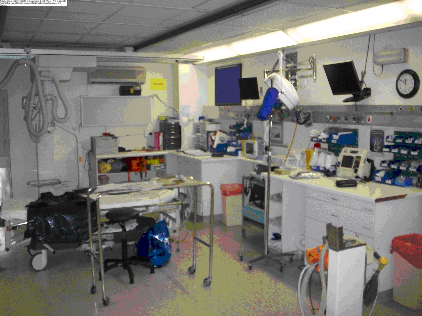
Trauma bay at Milbank Hospital (Johannesburg, South Africa).  This is a private hospital that caters to those who can afford private medical insurance and foreigners.


They have told me stories of aggressive surgeons barging into a blood bank and taking a unit for transfusion without appreciating that the ABO group did not match and of nurses hanging a bag of blood for a child, transfusing half the unit, putting it back in the refrigerator in non-sterile conditions, and then transfusing the rest of the unit to another patient later in the week. Both resulted in fatalities. Developing programs to educate clinicians could save numerous lives.

The first stop on my tour was the South African National Blood Service (SANBS). This organization has resources and expertise atypical of the rest of the continent. The medical director is a board certified internist trained in Washington. Their standard of care is more similar to what we offer in the US. They perform serological and nucleic acid testing for HIV, HBV, and HCV on all donors, reducing the risk of transmission to about 1:400,000 transfusions. (We advertise rates of 1:2,000,000 at the Brigham with our procedures; however, we don�t have a HIV prevalence of 20% in our general population.) They separate blood components and provide advice to clinicians across the country regarding transfusion reactions and blood products.

I was able to watch their testing and component processing and compare it to our procedures at BWH � all were very similar except their equipment was slightly older. Their physicians also remotely oversee therapeutic apheresis and stem cell collections, which will be driven back to their main offices, processed, and driven to the recipient�s hospital for infusion. I observed the apheresis clinics and again found their equipment and protocols almost identical to what we use at BWH. However, I am assured that the standard available in South Africa and Johannesburg in particular is not typical of the rest of the continent. I am excited to find out for myself �.

Therapeutic apheresis clinic in Auckland Park (Gauteng) South Africa.  We had just finished a hematopoietic stem cell collection.

Implementation of basic oral health care delivery systems in Haiti (Deux)

Cap Haitien, Haiti

Today we packed our supplies and equipment and headed to the clinic in the Justinian University Hospital about 10 blocks away.  The Justinian is the main public hospital in town with 250 beds serving almost a million people in northern Haiti. 
At the oral maxillo-facial surgery/dental clinic in the Justinian, my site mentor had set up an educational day session where we train local Haitian dentists and nurses in restorative techniques practiced in the States.  We dragged in box after box of materials, equipment, and supplies and eagerly dove into seeing the waiting room full of patients.  It didn't take long for the day to take a downward turn.  Let me explain.  Modern dentistry in America is a smorgasbord of products, with every procedure requiring specific instruments and materials.  You want a simple clear filling?  Of course, just go get the high speed drill, low speed drill, 330 bur, 245 bur, #6 round bur, 2% lidocaine with 1:100,000 epinephrine, 20% topical benzocaine, local anesthetic syringe, 27 and 30 gauge needles, cotton tips, 2x2 gauze, patient bibs, high speed suction, low speed suction, air/water syringe, mirror, explorer, endo ice, barrier tape, Fuji liner/base, mixing spatula, mixing pad, curing light, phosphoric acid etch, Optibond Solo, applicator tip, A3 Herculite composite, plastic instrument, Mylar strip, wedge, composite finishing burs both flame shape and football, college pliers... and yes, this is the basic setup for one restoration.  All these materials need to be laid out and prepared before the actual procedure due to the setting times of the materials resulting in a race against the clock.  Imagine the flurry of activity in the small, two-chaired room as we realized mid-treatment that we were lacking essential instruments and materials in the very specific procedures we were performing.  This was complicated by the malfunction of the overhead lights, compressor breakdowns, and a flood of constantly leaking water, which resulted in moisture leakage in restorations requiring dry fields.  A primitive "sterilization room" was set up across the hall, but mostly we wiped down handpieces et al with extra masks and a dollop of Purell.  In addition, most of the restorations we performed were heroic efforts to save what remaining structure was left in symptomatic patients who essentially needed more complicated procedures but could not due to time, supply/equipment limitation, and finances.
Facial trauma also fell under the auspices of the clinic, as seen in the photo below of a woman 4 days s/p motorcycle accident.  The local dentist explained to us that in the past 3 years, motorcycle usage and thus accidents had increased exponentially in Cap Haitien.  For this patient with a through-and-through philtrum/upper lip and infraorbital laceration, only one interrupted 1-0 nylon suture was available to close the lac.  Having just come from rotation at MGH oral maxillo-facial surgery, it was a different world to say the least.
In addition to the mechanical complications, the dynamic between our team and the local health care providers had to be handled with utmost delicacy, as we were careful to emphasize a symbiotic learning relationship instead of imposing a foreign "know-how" attitude.  At the end of the day, all the biohazard waste was dumped outside next to the steps of the clinic.  Our team looked at each other, defeated.
We had a long discussion with our site mentor, who revealed that this day was constructed to reveal the gaps in our approach to public health, specifically when we apply our American protocols to a situation that cannot be translated in Haiti.  Despite our best intentions, a 30-piece setup with reliance on dependable water, electricity, high technology, and sterile fields are not completely reasonable nor at times appropriate.  Sometimes it is necessary to forgo our strict tutelage and employ a more practical philosophy.  It is the unspoken rule in Haitian healthcare- rules are meant to be broken.
This was further emphasized during dinner with a local urologist, who told us stories of water leaking from the ceiling into his sterile field in his ORs, making molds for amputees' leg prosthesis from 2-liter coke bottles, and chickens running through the surgical wards of the hospital.  He told us how after 20 years of work in Haiti, he leaves every trip questioning his contribution and purpose.  It is after days like this and conversations like this that make me understand Haiti and the draw of Haiti more- that this piece of land with its complicated history, politics, and tensions makes it into the Rubik's cube of the public health universe.

Implementation of basic oral health care delivery systems in Haiti (Un)

Cap Haitien, Haiti

Bon jou, s'ak passe, salut.  There are many ways to greet someone in Haiti, a true m�lange of cultures and opinions in one small but electric country.  Haiti has been a "hot topic" in a variety of forums- international media, politics, and of course inside the walls of the Brigham.  As a longtime Partners in Health fan and global health enthusiast, my goal is to investigate the process of implementing an oral health delivery system in Haiti that is sustainable, appropriate, and affordable to deliver.  Using the World Health Organization guidelines as set forth in the highly under-used Basic Package of Oral Care as our model, we hope to merge education and direct care in one program that integrates the local population but can be applied globally.  Additional aims of this directive are as follows:
Ensure that the oral health care provisions established are sustainable by training local Haitian nurses to be dental assistants.
Implement and utilize the Atraumatic Restorative Treatment method, which allows for restoration of dental decay in basic accomodations without electricity or water-dependent equipment.
Provide direct oral health care to Haitian residents including emergency relief, preventive care, and comprehensive dental services.
To establish a continuing education, referral, and teaching center for the North Haiti Dental Association (NHDA) doctors.
Vision and supplies were packed into checked luggage and I set forth to Port-au-Prince, a maze of rebuilding and tent cities slowly regenerating two years after the devastating 2010 earthquake complicated by lack of infrastructure and a cholera outbreak.
Port-au-Prince: Cholera public service announcement found on most public buildings in Haiti.  From the capital, we flew on a verified joyride to our site in Cap Haitien on a small 15-seater over the mountain ranges Massif du Nord and Montagne Noires.
Re-usable airline ticket to Cap Haitien.  The airport consisted of a one-room terminal.  The waiting room for the airport was outdoors.  We were transported to and from the airport in the bed of a pick-up truck, which was actually a delightful experience.
I was told by a local source that there are approximately 10,000 NGOs in Haiti, with a good portion operating in Cap Haitien, a large city nestled between mountains and sea in the northeast of Haiti.  We were de-briefed on our program and schedule of visiting several clinical sites in addition to various didactic lectures. 
From my time in Cap Haitien thus far, I have made a few observations about this dynamic culture.  One, there is a palpable charisma throughout the entire place that understandably attracts the international community.  Two, there are no trash receptacles to be found, and every type of disposable waste ends up on the street, in the ocean, or on the beach [picture below].  The grime on the streets is in stark contrast to the dignified manner and careful grooming of the Haitian people, best visualized in church, which is well-attended.  Religion is engrained in the core of the culture; when driving down the street one passes "Lord the Savior Auto Repair" and "Jesus Christ the Good Shepherd Barbershop."  Walking down the circus of dirt roads, you hear the roar of motorcycles narrowly dodging pedestrians, the smell of petrol and burning, and the coating of dust all synthesized together in a term my site mentor coined as "a potpourri of poverty."  However, I have seen such developmental potential in Haiti as we passed by miles of pristine beaches, leafy mountains, and quaint landscape ripe for economic opportunity.  The situation here is truly unlike any other that I have seen in previous global health experiences in Nicaragua, Bolivia, China, and Botswana.  I am both intrigued and energized to immerse myself in this clinical and educational experience.

An American Pathologist in Malawi: A Tale of Two Cities

Kevin Golden, MD/PhD
PGY-5, Surgical Pathology Fellow

The infrastructure for most of Malawi�s pathology is set in the country�s two largest and most populous cities: Blantyre and the capital, Lilongwe.  They are separated by 366 kilometers of mud-soaked highway, speckled with farming villages and local markets along the entire route.  This translates to about a four and half hour bus ride, which doesn�t seem like a long length of time to squeeze into the seat, unless that person (me) was six foot five.  Nonetheless, outside of my leg falling asleep and the bus having to swerve around the occasional stray goat in the road, our journey was an uneventful one. 

Pathology laboratory in Lilongwe
The hospital in Lilongwe is set at the top of a hill overlooking part of the city.  We, as pathologists, don�t have a large amount of daily exposure to patients, so our tour started and ended in the pathology laboratory.  I entered the lab with similar expectations as to what I had seen while working for the last two weeks in the College of Medicine in Blantyre.  I quickly found out that this was not the case, however, as this pathology lab looked something like out of a catalog: clean, shiny metal workbenches with all new automated equipment, imaging software attached to the computer, and, as spoiled as this sounds, air conditioning.  (Although the open-air pathology sign out room in Blantyre has a nice cross breeze in the afternoons, there is something about a cool 73 degrees that allows one to have better focus on the task at hand.) 
All of these factors seemed to improve the quality of the H&E stains on the slides, even to that which is comparable to what I see back in Boston.


Unfortunately, the challenges of diagnosis remain the same.  The specimens are usually accompanied by a limited clinical history and no available ancillary immunohistochemistry tests are available.  There are also several similarities in the surgical samples, which include some of the largest and most destructive tumors I have seen in my short career as a pathologist.  I�m not quite sure if the reason that patients wait to see a physician is more of a national infrastructure problem or a problem of access for care, but I digress. 

Reviewing cases in Lilongwe
We stayed in Lilongwe for two days, cutting in surgical specimens from the prior week, and diagnosing cases that had been submitted as long back as November.  There are no full-time pathologists in Lilongwe, so cases are compiled together either to be read every couple of weeks by a visiting pathologist or sent down to Blantyre to be read by the full-time pathologists stationed there.  Either way, the turn around time is a week or two at best.  At its worst, it can be months.

Despite differing outside appearances for both locations, the problem remains the same: too many specimens and not enough staff.  The entire country of 22 million people employs three residing full-time pathologists.  These pathologists are responsible for signing out cases, as well as teaching and administrative duties.  The logjam of cases can cause a delay in reporting results, thus causing a delay in crucial treatment.  This is where I feel that my work as volunteer pathologist becomes most important.  I hope that in my short time I can indeed make a difference, no matter in which city I am.

An American Pathologist in Malawi: Establishing a Baseline

           It seems that every specialty in medicine needs a baseline in which to compare.  Laboratory values, CT scans, nutritional intake, even mental status, all need a point of reference to which it can be compared to the baseline.  I can still remember back when I was a medical student on the wards, learning that certain patients were consistently outside of the �normal� values, and that there was no cause for concern, all attributed to the patient�s baseline.  It�s a funny thing, as each and every patient has a different baseline, and we, as clinicians, need to be able to gauge each one in the context of that patient.   What factor may be remarkably noticeable in one patient may be completely within the realm of �normal� for another.  The same goes for pathology, as strange as that may sound.  It is easy to think that a specimen is simply a specimen and slide is nothing more than a slide, and there should be no difference.  I must admit that I had that same line of thinking before arriving in Blantyre, Malawi.  While rotating on various BWH specialty consult pathology services, we as residents always have seen a wide variation of quality from outside hospitals, ranging from the gross description of the specimen to the H&E staining to the immunohistochemical methods used to come to the final diagnosis.  During signout of these cases, the pathology attending and I will usually discuss all of these aspects, and how fortunate we are at BWH to have consistent quality for processing the cases.  We constantly had to keep in mind that the cases from each of the different laboratories had a different baseline, of which we needed to be able to interpret accurately.


            With this mentality, I traveled to Blantyre fully expecting crystal-clear gross descriptions of specimens, as well as flawless H&E staining and an arsenal of immunohistochemical stains that would allow me to make the diagnosis with relative ease.  Much to my surprise (and naivety), my orientation to the pathology lab was not what I was expecting, or in my comfort zone.  My departmental tour started out like any other, having the program director show me the administration office and labs.  I found out that the department was in the process of switching to electronic records, so any prior pathology reports I would need were awaiting me in a neatly stacked pile of binders.  No big deal, I thought, as all the reports looked quite organized on those shelves, and this was their baseline for record keeping.  My tour continued through the lab, where I was shown where the specimens were cut and processed.  The space, which included an open-air view of the grassy quadrangle, was certainly adequate for the job that needed to be done.  I was introduced to an extremely friendly member of the laboratory, Kingsley, who was there cutting the paraffin blocks for slides.  I originally thought that he was the laboratory technician, but I discovered later that he is a board-certified cytopathologist that reads all of the Pap smears that come into the department. 

As I looked around, I couldn�t help but notice that there were no immunohistochemistry technicians.  Back at BWH, the immunohistochemistry lab for pathology consists of five full-time technicians processing over 200 custom orders each day.  It was at this point that is was casually mentioned to me that there was no immunohistochemistry lab.  No immunohistochemistry lab?  For all the times at BWH that I would settle in the comfort that any questionable issues (big or small) I had with a case could most likely be solved with immunohistochemistry.  That invisible crutch was suddenly taken away, and I had been thrust into a realm of relative insecurity.  What do you mean I can�t confirm this obvious squamous cell carcinoma with a p63 stain?  This department in Malawi had been diagnosing pathology cases long before I got there, without the help of immunohistochemistry.  This was the baseline in Malawi, and diagnoses had to be made, independent on ancillary studies.  I realized that this experience was going to force me to establish my new baseline, and I eagerly anticipated the challenge.  

I was left to the basics: hematoxylin and eosin stains and my armament of knowledge of medical school histology and my resident training in pathology.  Even after this initial shock, I remained confident that I would be able to handle the situation as I was subsequently shown to my workstation.  I was assigned to a nice multi-headed Leica microscope (the only multi-headed microscope in the department) and given my workload for the day.  This will be a challenge, I thought, as I placed the first slide down on the microscope stage.  Gasp.  There must be something wrong with this scope!  The H&E stain was faint, and nothing like the vibrant colors that I had become acquainted with back at BWH.  It�s as if someone had turned the contrast and intensity down on the slide using Photoshop.  For a while, I played with the focus, light intensity and condenser in a futile attempt at improving my perspective, ultimately realizing that the colors I was seeing were just another baseline that I would need to establish. 

The diagnoses were certainly there in front of me, and I was going to be forced to retrain my eye to be able to pick up these subtleties in order to make my correct diagnoses.  I�ve been presented with these sorts of challenges before, and I was confident that my prior training would serve me well.  After about five slides, my heartbeat slowed to a regular rhythm (back to its baseline) and I settled into my comfort zone, identifying familiar entities that I had seen back at BWH.  I had established my new baseline, and the anxiety of being outside of my comfort zone quickly subsided, as I continued through my cases for the day.

Kevin Golden, MD/PhD
PGY-5, Surgical Pathology Fellow

Scaling up head trauma and critical care capacity in a resource-poor setting: Mbarara Hospital, Uganda (4 of 4)


Thursday, November 10, 2011

Today I was finally able to demonstrate the drill to Gerald and the residents. Since there had been no appropriate cases during the week, we bought some fresh goat heads from the market and performed burr holes and craniotomies on them. Fortunately, the electricity cooperated. The residents had a great time learning to put the drill together, practicing with the different attachments, and working on the goat heads. Among other things, we discovered that a goat�s frontal bone is very thick under its horns! We also discussed basic head trauma management, and the residents showed an excellent grasp of the relevant anatomy, physiology, and critical care despite having few opportunities to practice it. I reviewed the technique of exploratory burr holes, which I have never actually performed � because CT and angiography have been widely available for so long, few if any practicing neurosurgeons in the US have performed this technique. The residents helped me get a sense of what they try to do for head trauma patients in the ER. Since imaging is not available, they rely on the history and exam. Any loss of consciousness warrants 24-hour observation. They use the Glasgow Coma Scale, vital signs, and pupillary asymmetry to decide whether to give mannitol (intermittently available in the ER) or explore. However, even at MGH, by the time a patient exhibits the classic Cushing response or �blows� a pupil, it may be too late. Also, once a patient is admitted to the ward, such monitoring no longer occurs. It is common for patients to come in with a GCS of 13 or 14, then suddenly decompensate and die on the ward � presumably because of a rapidly expanding epidural or subdural hematoma that could have been addressed if an accurate imaging diagnosis had been available.

Through this discussion, I gained a much better understanding of the situation in Mbarara. This urban hospital is caring for head trauma patients every day, and the doctors have an excellent knowledge base � but they lack the resources to effectively triage and monitor patients. My sense is that the primary obstacle is inadequate monitoring on the ward - ongoing assessment of mental status, hemodynamics, chemistries, etc. � things that we might expect to occur in an ICU or stepdown unit. Thus, scaling up ICU care in Mbarara might go a long way toward improving head trauma outcomes. It will also be crucially important to have a CT scanner in order to diagnose intracranial hemorrhage in patients who have not already decompensated. But even without imaging, ongoing assessment of clinical exam will help capture more patients who require neurosurgical intervention.

Although I was only in Mbarara for a short time, I plan to return in the next few months � and to establish ongoing collaboration between my department and the surgeons there. Since they are used to a high-cost, high-complexity system, neurosurgeons in the US often take a fatalistic attitude toward global health, believing they cannot possibly contribute. I hope to help demonstrate the opposite � that neurosurgeons can and should help build up local resources to care for head trauma patients, reducing its worldwide burden of death and disability.

Anna R. Terry, MD, MPH
PGY-5, MGH Department of Neurosurgery

Like Us

Blog Archive