Medical News Blog Information

Editor's comment: Biomolecular Detection and Quantification...

I've been invited to be a section Editor for Biomolecular Detection and Quantification (BDQ).

BDQ is an Elsevier-based, open access, peer-reviewed journal dedicated to championing excellence in molecular study design, measurement, data analysis and reporting. 


BDQ's Editor's-in-Chief are Stephen Bustin, Jim Huggett, Justin O'Grady, Michael Pfaffl, Carl Witwer and Ron Cook. Quite the line up huh?


My role will be one of attracting, reviewing and shepherding manuscripts relating to BDQ in infectious disease, but particularly related to viruses.

Our two main aims
are:
  1. to provide a forum for discussion and recommendation of guidelines designed to improve the accuracy of molecular measurement, its data analysis and the transparency of its subsequent reporting;
  2. to publish molecular biology based studies that adhere to best practice guidelines, both current and future.
So, if you have any ideas for future articles or would like to publish in our little baby right now (once they become toddlers things get more rowdy), please contact me or one of the other Editors as soon as you can.

Links...

Editor's Note #14: VDU in the scientific literature

Virology Down Under gets a 2nd citation (1)!

Dr Anne S De Groot and colleagues used a VDU graphic for a recent paper and cited VDU for it. Many thanks!

To read the paper entitled Cross-conservation of T-cell epitopes: Now even more relevant to (H7N9) influenza vaccine design hop over to..

  1. 1st citation for VDU at http://newsmedicalnet.blogspot.com.au/2013/10/concise-overview-of-mers-from-mid-2013.html

Snapdate: Middle East respiratory syndrome coronavirus (MERS-CoV)

Click on image to enlarge.
Chart update time for human cases of the MERS-CoV.

Chart #1: lab confirmed(Ministry announced) human cases of MERS by region of acquisition. MERS is a disease with its activity firmly rooted in Saudi Arabia.

Chart #2: age and sex distribution pyramid. MERS is a disease of older males.


Click on image to enlarge.
Chart #3: here we see total and just the fatal MERS-CoV cases in humans, by month and year. I've kept the same value on the y-axis (left side) for ease of comparison. This chart highlights that there is no obvious seasonality for cases. In 2014 we're seeing more in Jan and Feb than we did in 2013 and in 2013 cases trickled along at similar levels from April onwards. This, if anything, supports the sporadic acquisition of cases and rejects the idea that, at least among the hospital-based population that is the main one to have been tested to date, MERS-CoV is not a seasonal virus.


Click on image to enlarge.
Chart #4: this one shows as the slowly spreading outbreak. More disturbingly, it shows the very gradually increasing PFC. Currently, 43% of MERS cases have died; 2 in 5. Guess its worth remembering that MERS-CoV may a puny virus in camels and in terms of transmission efficiency; but not at all puny in regards to what it does to the older usually male and mostly already ill people it infects. It is quite an efficiently lethal virus in that sub-population.

Coming back to MERSerable data...

A grab of the past 20 MERS-CoV positive human cases reported
by a Ministry and listed over FluTracker's if you want to look in
more detail. Even though the tally goes to 191 - there are 186 total
cases
(some have been removed after being reduced in status to
probable cases). Under the FluTracker Case # banner, a pink fill
indicates a death. A pale blue fill under the Sex column indicates that
the World  Health Organisation (WHO) have reported the cases through
their Global (GAR) Disease Outbreak Notice (DON) website.
Click on image to enlarge.
I'm preparing a brief invited talk on MERS and MERS-CoV for next week at the Molecular Microbiology Meeting in Sydney

So I'm updating MERS-CoV data - 4 cases since last I did this. And the data remain as horrible as the last time I complained

The figure above highlights just how horrible. Have a look at:

  • How many data gaps there are for sex (a very basic piece of demographic information to provide without comprising patient identity)
  • How many data gaps there are for date of illness onset
  • The absence of any KSA unique identification codes (there were 4 provided in August...and that was that)
  • How many dates of hospitalisation there are
I'll throw up some new charts shortly but really, they will reflect these data gaps.

But really, this has all been said before so I won't rehash my disappointment too much. 

Except to say..

It isn't at all surprising to read a comment like that from Prof Ian Lipkin recently. This being in the context of his unreciprocated collaboration with the Kingdom's Deputy Minister of Health, Dr Ziad Memish...
We've gone our separate ways, and I wish him well
...which may also inform us about why the collaboration between the MOH and the World Health Organisation (WHO) produces such spartan data on MERS cases, at least when you compare the quality of data to that which China provide WHO on avian influenza A(H7N9 virus cases; a much larger undertaking involving a more populous State and many more geographic and political boundaries.

Vale Dr. Albert Z Kapikian

Others will no doubt write more cogently and personally than I can about Dr Kapikian's >56-years of medical and virology expertise, skills, discoveries and personality but I wanted to make a just a few quick comments on his passing in relation to a group of viruses for which he is not as often associated; the rhinoviruses (RV).

Dr Kapikian was an expert with electron microscopy (EM), learning the use of it from June Almeida at the Royal Postgraduate Medical School, University of London in 1970. His use of immune EM led him to discover the viruses we now call noroviruses in 1972; a major cause of gastroenteritis. He also found instances of rotavirus (RoV) in the United States and went on to lead the development of an oral vaccine to prevent the serious diarrhoeal disease associated with infection by RoVs. 


He has over 200 publications listed on PubMED.

I exchanged a couple of eMails with him in 2009 (which Al graciously replied too in excellent detail) but never got to meet him, much to my disappointment. My first knowledge of him was from my work with rhinoviruses. While these were not his research focus, he Chaired the group that published the 1st (and 2nd in 1972 and co-authored the 3rd in 1987) formal description in 1967. This report gave the largest group of distinct respiratory viruses their 100 names. He helped to bring together RV researchers and their often identical isolates from around the globe and helped to create the 1 key to link them all in a single unifying scheme; HRV-1 to HRV-100. Having had a tiny role in doing that for the latest species, RV-C, I can more than imagine how much of a challenge that must have been at the time. Dr Kapikian also used his immune EM expertise to visualize the rhinoviruses in 1972.


That Dr Kapikian is so highly regarded, described as warm-hearted and the smartest and nicest guy in the room, speak volumes of him as a human. His track speaks to his role as a leader and medical virologist of huge impact.


Another trailblazer has moved on.


Dr Albert Z. Kapikian, Chief, Epidemiology Section, Laboratory of Infectious Diseases, National Institute of Allergy and Infectious Diseases, National Institutes of Health in the United States, died 24-Feb at 86-years of age.


References...

  1. National Institutes of Health statement on the death of Dr Albert Z Kapikian
    http://www.niaid.nih.gov/news/newsreleases/2014/Pages/AlbertKapikian.aspx
  2. Seeing the new calicivirus: norovirus
    http://jvi.asm.org/content/10/5/1075.long
  3. Context for the discovery of norovirus
    http://www.ncbi.nlm.nih.gov/pubmed/?term=kapikian+a+s295+norwalk
  4. Sabin Gold Medal
    http://www.sabin.org/sites/sabin.org/files/GoldMedalSpeech2005.pdf
  5. Dr Kapikian's publication list on PubMED
    http://www.ncbi.nlm.nih.gov/entrez/query.fcgi?cmd=search&db=pubmed&term=%28kapikian%20az%5bauth%5d%29
  6. NIH on the Sabin medal
    http://www.nih.gov/news/pr/may2005/niaida-12.htm
  7. Children's Vaccine Initiative Pasteur award summary at NIH
    http://nih.gov/news/pr/nov98/niaid-10.htm
  8. Alumni Award of Distinction from Weill Cornell Medical College where Dr Kapikian got his MD
    http://weill.cornell.edu/news/news/2001/06/kapikian-and-szeto-receive-alumni-awards.html
  9. Awarded Fellowship of The American Academy of Microbiology
    http://nihrecord.od.nih.gov/newsletters/2012/06_25_2012/story6.htm
  10. Visualising rhinoviruses by immune electron microscopy
    http://jvi.asm.org/content/10/1/142.long
  11. First rhinovirus numbering system report
    http://www.nature.com/nature/journal/v213/n5078/pdf/213761a0.pdf
  12. A collaborative report: rhinoviruses--extension of the numbering system from 89 to 100.
    http://www.ncbi.nlm.nih.gov/pubmed/3037780
  13. A collaborative report: Rhinoviruses-extension of the numbering system
    http://www.sciencedirect.com/science/article/pii/0042682271903291

Dromedary camels are a host of MERS-CoV...

Yes. Not a "MERS-CoV-like" virus or "something very closely related to but slightly different" from MERS-CoV. Camels. Are. A. Host. 

There was already plenty of evidence to suggest this (see some of my previous posts on this linked below), and none to really dissuade me from thinking otherwise. And yesterday we saw a new paper by Ian Lipkin and his collaborating crew from King Saud University in Saudi Arabia that make this issue more obvious than ever. 


So let's stop messing around. There is an elephant in the MERS-room...and its a camel! 


The Middle East respiratory syndrome coronavirus, does in fact look to be a camel virus that causes few symptoms in that host, is acquired by young camels and has been for at least 22-years, and then people somehow get infected, probably from proximity to camels or due to habits involving camels. the keeping of camels or at gatherings in which grumpy slavering camels are congregating. 


Yes, there is little evidence for any contact with camels among the 186 human cases as Dr Ziad Memish, deputy health minister of the Kingdom of Saudi Arabia (KSA) points out, but as Prof Marion Koopmans noted to NPR"few people [with MERS] have had the kind of follow-up you would want". So we take denial of any contact with a grain of salt.


There is definitely some contact however; some that is pretty solid. For example, the owner of a camel in Jeddah (4) that both tested positive for MERS-CoV or the Qatari farm camels and owner and an employee that were all MERS-CoV positive (5,7). 


And there is far less evidence against camels as a source of  some/many/most human cases and for anything else. 


So in the new paper in mBio published online 25-Feb, we read of the most comprehensive KSA camel study to date. Camels from 2013 were sampled and camel sera collected and frozen since 1992-2010 were tested and many were found to be MERS-CoV antibody positive. From the more recent dromedary camels, nasal swabs were also found to be viral RNA positive; RNA that is definitely from MERS-CoV.


Some key findings...


  • No sheep or goats were MERS-CoV antibody or RNA positive; a routine finding now. Bovine CoV antibody reactivity was identified in these animals however, and in 17% of camels
  • Antibody was detected using infected cells and also using a method that employs a specific portion of a MERS-CoV protein (part of the nucleoprotein)
  • 150/203 (74%) of camels from all over the KSA had MERS-CoV antibodies in a pattern reminiscent of any endemic human respiratory virus
    • 95% in camels older then 2-years (adults)
    • 55% in those =2-years (juveniles)
    • The south west had the lowest proportion of positive camels (5%)
    • Higher proportions were found in central KSA (Riyadh)
  • 3 rectal swabs were positive for MERS-CoV RNA using real-time reverse transcriptase polymerase chain reaction (RT-rtPCR). 2/3 camels were also nasal swab POS
    • 36/104 juvenile camels were nasal swab POS
    • 15/98 adult camels were POS
    • 66% of samples from the west (Taif) were POS but none from the south west
    • no RNA was detected in a sampling of camel blood/sera and so the archived samples from earlier years could not be sequenced to verify that they had MERS-CoV sequences in them
  • Amplification and sequencing of a 1,044nt portion of the Spike gene, 2,004nt ORF1ab region found that less than 1% difference from previous published MERS-CoV sequences and the nucleocapsid gene region was identical. Great to see a move away from recent reliance on complete genome sequences and a more practical and rapid subgenomic, multi-target molecular epidemiology approach used. 
    • 11/13 higher viral load samples could be amplified and sequenced
It was interesting hear the TWiV (this week in vrilgy) podcast interview with Prof Lipkin and Assoc. Prof Thomas Briese in which they noted:

  • MERS-CoV is a "puny" virus causing little overt disease on camels
  • The MERS-CoV genome seems to be fairly stable; its not influenza virus and does not seem likely to evolve rapidly
  • Baboons, dogs, cats, rodents are on the list to test when the team return to KSA
  • There has been a previous report on limited human antibody levels to MERS-CoV in at least the east of the KSA


Sources and previous posts on camels and MERS-CoV...

  1. mBio paper by Alagaili and colleagues
    http://mbio.asm.org/content/5/2/e00884-14.full.pdf+html
  2. NPR's Richard Knox: excellent story
    http://www.npr.org/blogs/health/2014/02/25/282136478/deadly-mers-virus-circulates-among-arabian-camels
  3. CIDRAP Story
    http://www.cidrap.umn.edu/news-perspective/2014/02/study-mers-cov-may-have-been-saudi-camels-22-years-ago
  4. Camels owner in Jeddah
    http://newsmedicalnet.blogspot.com.au/2013/11/camel-cough-coronavirus-caught.html
  5. Two Eurosurveillance studies reporting MERS-CoV antibodies in camels
    http://newsmedicalnet.blogspot.com.au/2013/12/middle-east-respiratory-syndrome.html
  6. MERS-CoV antibodies in 10-year old UAE camel sera
    http://newsmedicalnet.blogspot.com.au/2014/01/antibodies-in-10-year-old-uae-camel.html
  7. More on the Qatari camels and some MERS-CoV sequencing and social media chatting
    http://newsmedicalnet.blogspot.com.au/2013/11/dutch-researchers-in-collaboraion-with.html
  8. MERS-CoV antibodies in camels from the Canary islands and Oman
    http://newsmedicalnet.blogspot.com.au/2013/08/camels-carry-signs-of-coronavirus.html
  9. Early cautionary thoughts from the WHO
    http://newsmedicalnet.blogspot.com.au/2013/08/who-urges-dont-put-camel-before-cart.html
  10. Thoughts about MERS-CoV acquisition
    http://newsmedicalnet.blogspot.com.au/2013/09/most-mers-may-not-have-met-camel-but.html
  11. Querying whether there is a better possible source for human cases
    http://newsmedicalnet.blogspot.com.au/2013/09/is-there-better-smoking-bat-or-camel.html
  12. Summing up the first 100-days of (human) MERS-CoV infections
    http://newsmedicalnet.blogspot.com.au/2014/02/middle-east-respiratory-syndrome.html
  13. MERS-CoV antibodies in camel sera dating back to 2005 in the UAE
    http://newsmedicalnet.blogspot.com.au/2014/01/mers-cov-antibodies-in-dromedary-camels.html
  14. Gatherings and acquisition/transmission of MERS-CoV between animals and humans
    http://newsmedicalnet.blogspot.com.au/2014/01/a-date-with-middle-east-respiratory.html
  15. Qatari camels clear the MERS-CoV from their systems
    http://newsmedicalnet.blogspot.com.au/2013/12/qatari-camels-clear-coronavirus.html
  16. TWiV podcast
    http://www.twiv.tv/2014/02/25/twiv-special-mers-coronavirus-in-dromedary-camels/

Using human volunteers to understand influenza virus behaviour and its impact....

This will be the first in a long term project to catalogue the literature's record of volunteer infection studies using influenza virus (IFV). Much like the one I started on rhinovirus transmission back in Nov-2013...see those posts here and here, so far.

I don't guarantee it will be done before I'm too old to type, but I'll be adding to it as time allows. ;)


So first up is a paper.

Author: Frederick Hayden et al
Journal: J Clin Invest 101(3):643-9
Year: 1998
IFV strain used: A/Texas/36/91 (H1N1)
Inoculation route/amount/viral titre: intranasal/0.25ml/105 TCID50

Volunteers included: 19 healthy susceptible males (26%) and females, median age 21-year
Antibody levels: =1:8, haemagglutinin-inhibition (IH)

Some key results...


  • Volunteers were kept in isolation for 1-day prior to inoculation.
  • Most (74%) volunteers shed virus on day 1 (1 day after inoculation) as determined by viral growth from recovered nasal washings
  • All volunteers developed symptoms with scores peaking at day-2 and returned to normal by day-8
  • 12/19 (63%) developed fever, peaking at day-2
  • Upper respiratory tract signs of illness (runny nose, sore throat occurred in 15/19 (79%), peaking at day-2
  • Lower respiratory tract symptoms (cough, hoarseness) peaked on day-5
  • Systemic symptoms (muscle aches, fatigue) also peaked on day-2
  • Proinflammatory cytokines in nasal washes revealed several patterns:
    1. Interleukin (IL)-6 and interferon (IFN)-a increased (day-2), decreased, then increased again (day-5) - a biphasic pattern
    2.  IL-8 tumour necrosis factor (TNF)-a began rising at day-2, peaking at day-4; IL-8 (peaking when nasal mucous turbidity increased) not decreasing as rapidly as TNF-a
    3. IL-1�, IL-2 and tissue growth factor (TGF)-� did not rise
  • Cytokines in plasma and serum
    • IL-6 and TNF-a peaked weakly but at the same times as in the nose 
    • IFN-a and IL-8 were not detectable
    • IL-1�IL-2 and TGF-� did not rise
  • Nasal lavage virus levels (titres) correlated with fever, total symptoms, systemic symptoms and upper respiratory symptoms, TNF-a, IFN-a and IL-6 on day-2. Total symptoms, viral titre, IL-6 and TNF-a all correlated on day-5
  • Lower respiratory tract symptom scores peaked in correlation with IFN-a and IL-6 on day-5 & day-6 (also when IL-8 peaked)
As you'd expect given the inoculation route, signs and symptoms of disease occurred first in the upper respiratory tract and then progressed to the lower respiratory tract over time. This is obviously not a "natural" infection as we do not get 0.25mL of flu-containing liquid flying up our noses when someone sneezes on/near us. So the value of this study lies other than in studying natural transmission.

The authors note a few things in the discussion...


  • The response to infection at the site likely leads to the local (runny nose,cough) and the systemic (fever, myalgia) signs and symptoms. 
  • Cytokines are produced by a variety of cells; monocytes/macrophages, bronchial epithelial cells, CD4+ and CD8+ T cells and the infected epithelial cells themselves.
  • Mouse models seem to have more severe lower respiratory tract disease after influenza virus infection than do human volunteers
  • Other cytokines may be involved during more severe natural influenza infections of humans
  • TNF-a, although rising earlier, peaked later perhaps acting to control inflammation after virus has been contained (in the upper respiratory tract at least; what's happening in the lower respiratory tract?)
  • TNF-a and IL-8 were part of the 2nd wave of cytokines and may be better markers of more severe lower respiratory tract influenza disease
Cytokine background information...
  • IL-6
    • A proinflammatory cytokine
    • Secreted by T cells and macrophages
  • IL-8
    • Also known as neutrophil chemotactic factor
    • A chemokine secreted by macrophages and epithelial cells and anything with tool-like receptors
    • Attracts neutrophils and other granulocytes and indices phagocytosis in them
  • TNF-a
    • Also called cachexin or cachectin
    • Mostly secreted by macrophages,but also CD4+ lymphocytes and natural killer (NK) cells
    • It is a pyrogen, inducing fever, apoptotic cell death, cachexia (weight loss) and inflammation
  • IFN-a
    • A Type I interferon secreted by leukocytes, macrophages, epithelial cells, endothelial cells etc
    • Involved in the innate immune response against viral infection by triggering many other antiviral proteins and processes

Live bird market closures continue...

Changsha, capital of Hunan province has had its live poultry markets closed since 21-Feb (1).
H7N9 case map.

So that can be added to Shanghai, major markets in Hangzhou, Ningbo and Jinhua cities in Zhejiang (15), markets in Guangzhou, Guangdong ((3); plus a previous 2-week pause for disinfection in Shenzen (15) ), Hong Kong recently extended its ban (4) on live poultry imports from mainland China and Fujian provinces markets were closed fro disinfection back in 21-Jan. 

No word, that I can find anyway, on market closures in the Jiangsu province (11.8% of all H7N9 cases), other sites in Fujian province (5.5%) or Anhui province (2.5% but over-represented in cases recently).

And yet with no cases announced yesterday - the latest in a recent precipitous decline in total human cases per day - it's tempting to wonder whether market shutdowns in regions that have had the highest case activity (Zhejiang and Guangdong provinces) and which produce or trade a lot of the nation's chickens, have had enough of a flow-on effect to stem the tide in other regions supplied by these. 

Aside #1: Poultry eggs are mainly produced by >1-billion birds in Henan, Shandong, Hebei, Liaoning, Jiangsu, Sichuan, Hubei, Anhui, Heilongjiang and Jilin (7,8) whereas >4-billion broiler chickens (bred for meat) are more concetrated in Shandong, Jiangsu, Guangxi, Liaoning, Guangdong, Anhui, Sichuan and Henan provinces  (2,6,8,9,10,11,12,13).
Aside #2: An interesting to read (5,9) that the volume of chicken consumed per capita has risen by 9-fold or more in recent years - it has not always been the staple but is part of the tradition.

Sources...


  1. http://english.cri.cn/11354/2014/02/22/2702s814226.htm?utm_source=dlvr.it&utm_medium=twitter
  2. http://www.globaltimes.cn/content/843788.shtml#.Uwl4ovna6-1
  3. http://newsmedicalnet.blogspot.com.au/2014/01/zhejiang-live-bird-market-closures-and.html
  4. http://www.interaksyon.com/article/81029/hong-kong-extends-ban-on-live-poultry-imports-from-mainland-china
  5. http://www.iatp.org/documents/fair-or-fowl-industrialization-of-poultry-production-in-china
  6. http://www.forbes.com/sites/russellflannery/2013/04/13/by-the-numbers-chinas-poultry-industry/
  7. http://www.prweb.com/releases/chinas-egg-and-poultry/industry-research-2013/prweb11170628.htm
  8. http://www.ccagr.com/content/view/117/184/
  9. http://www.newschinamag.com/magazine/poisoned-plucked-processed
  10. http://www.tysonfoods.com/Around-the-World/International-Operations/Tyson-China/About-Tyson-China.aspx
  11. http://www.wantchinatimes.com/news-subclass-cnt.aspx?cid=1103&MainCatID=11&id=20140219000043
  12. http://articles.chicagotribune.com/2012-04-19/business/sns-rt-us-usa-china-food-factboxbre83j05v-20120419_1_hog-farm-province-hubei
  13. http://english.agri.gov.cn/hottopics/ah/201310/t20131028_20492.htm
  14. http://news.xinhuanet.com/english/photo/2014-01/22/c_133063832_2.htm
  15. http://news.xinhuanet.com/english/china/2014-01/31/c_133087279.htm

Thankfully we were also able to experience a little Vietnamese culture throughout the two weeks. I have posted several pics, including our visit to the Chu Chi Tunnels ( a major communist community that was a major battle ground during the Vietnam War). As you can see, we were able to explore the tunnels. It was a little claustrophic for sure!


Below are pics from our trip to the Mekong Delta, a small region south of Ho Chi Minh City, which is home to approximately 30 million people.


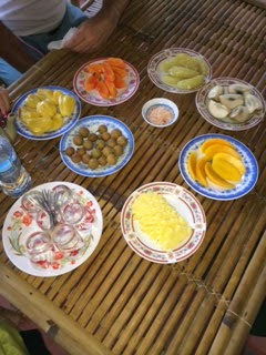
Vietnam is famous for it's vast selection of tropical, delicious, fruits. Truly the best fruit I have ever had.


Sadly, here is a pic of one of my final clinic days with Dr. Minh and Dr. Famy. This morning we saw 96 patients in a half day! Such a need for dermatology here. The work ethic of these physicians is just phenomenal.


2.17.2014

Greetings from Vietnam!

I have now been working with the brilliant dermatology faculty and residents of Ho Chi Minh City College of Pharmacy and Medicine for one week and I must say this has been one of the most memorable and impressionable learning experiences I have encountered throughout residency. 

A little background for you�.. For years, the country of Vietnam has lacked the appropriate means for effective, early intervention of disfiguring vascular anomalies.  The standard of care has historically included the use of radioactive phosphorus for infantile hemangiomas and even in some cases vascular malformations. This treatment is not only painful for the children but leaves behind disfiguring, stigmatizing, and painful scars, many of which are located in the facial region. Five years ago, Dr. Thanh Nga Tran and Thuy Phoung (both natives of Vietnman) decided they would put a stop to this treatment once and for all. Both having trained in the Harvard Dermatology and Dermatolopathology programs respectively, they determined to join forces with Dr. Rox Anderson (expert in the laser and medical treatment of vascular anomalies) and Dr. Martin Mihm (expert in vascular anomalies and dermatopathology).

Dr. Tran spent one month at the Ho Chi Minh College of Pharmacy and Medicine as a senior dermatology resident and during this time made contact with two of the most honest, hard-working, and caring dermatologists any of us has met, Drs. Hoang Minh and Bo Famy. Together, this remarkable team of physicians established the first vascular anomalies clinic of Vietnam five years ago this month.

The clinic began with 1 laser, the pulsed dye laser, an operating room with anesthesiologist, a team of eager residents, and of course a line of patients. Through much fund-raising and generous donations from various laser companies, the OR is now replete with the top four lasers needed for the treatment of various vascular anomalies. This accomplishment in only five years time is truly remarkable.

Now for the present�..Upon arriving in Vietnam at 1:30 AM, the hospitality of this program was evident immediately. Second year resident- Anh Dao was there with her little brother holding a sign with my name as soon as I exited the airport in Ho Chi Minh City. I must say this was so comforting having never been to this country and not speaking the language.

Our first day in clinic was truly an eye-opening experience. It began with the evaluation of several children with disfiguring scars from the treatment with radioactive phosphorus. In the year previously, Dr. Anderson was able to bring a new device that he invented which harvests a �blister graft� from the thigh, which can then be grafted to the site of a scar (after superficial epidermal ablation).  He trained Drs. Famy and Minh on the utility of this device and they were able to treat several of the children in the last year. The results are truly remarkable!  Nearly normal pigmentation and texture resulted. This is life changing for these children whose scars are located in the facial region. Additionally, this is ground breaking in the treatment of radiation injury. Thankfully, we were able to treat several additional children during our time this year.

Additionally, we evaluated and treated numerous children with hemangiomas, capillary malformations, lymphatic malformations, and venous malformations.
It was amazing to observe how brave these children are here. They literally walk into a room of 15-20 physicians and sit in the middle quietly while we discuss the treatment plan. 

A great deal of our time was also spent teaching the residents and attendings how to develop treatment plans, including the use of medical management, such as topical timolol and oral propranolol, in combination with laser treatment.  At the end of the week several members of our group spoke at the annual CME conference held at the University. Much to our surprise, this year�s attendance hit a record high of over 500 doctors from across Vietnam!


Like Us

Blog Archive